Lower Leg Bone Diagram / 1000+ images about Bones in the Leg on Pinterest | Bone ... - Human muscle system the muscles of the.. Lower jaw (mandible) collar bone. The humerus and the femur are corresponding bones of the arms and legs, respectively. There is also a knee cap called patella. The foot bones shown in this diagram are the talus, navicular, cuneiform, cuboid, metatarsals and hoof anatomy and bones of the lower leg. Ankle and foot bones and joints unit 4/12/18 lower leg:
It lies between the knee and the ankle while the upper leg lies between the hip and the kne. Posted on january 21, 2015 by admin. Ankle and foot bones and joints unit 4/12/18 lower leg: (2) hip bone attaches legs to our body. Start studying lower leg bone structure.
Bones Of The Leg Photograph by Asklepios Medical Atlas from images.fineartamerica.com 25.09.2018 · leg bone anatomy diagram diagram of human leg human anatomy diagram. The femur, or thigh bone, is the largest, heaviest, and strongest bone in the human body. The knee is a strong but flexible hinge joint. For more detail of the human bone structure, please visit: Start studying lower leg bone structure. Leg muscle drawing at getdrawings. The upper leg bone is connected to the lower leg bones at the knee by a hinge joint. The thigh bone, or femur, is the large upper leg bone that connects the lower leg bones (knee joint) to the pelvic bone (hip joint).
The humerus and the femur are corresponding bones of the arms and legs, respectively.
Download a free preview or high quality adobe illustrator ai, eps, pdf and high resolution jpeg versions. There is also a knee cap called patella. Several muscles attach to and act on the femur. At the microscopic level, this hard outer. Bones give your body structure and enable you to move, but what else is your skeletal system responsible for? Master leg and knee anatomy using our topic page. A leg bone is a bone found in the leg. Anterior view with primary bones names. The largest and most medial leg. Here's a diagram with the tibia bone labelled, as well as the fibula. Skeleton anatomical anatomy anterior view arm backbone biology board body bone bony chart chest diagram didactic education femur fibula finger foot graphic design hand health. (2) hip bone attaches legs to our body. Interactive tutorials about the lower limb bones, lower limb bones, os coxae, femur, patella, tibia, fibula, tarsal and foot bones, featuring images, diagrams and the beautiful illustrations of getbodysmart.
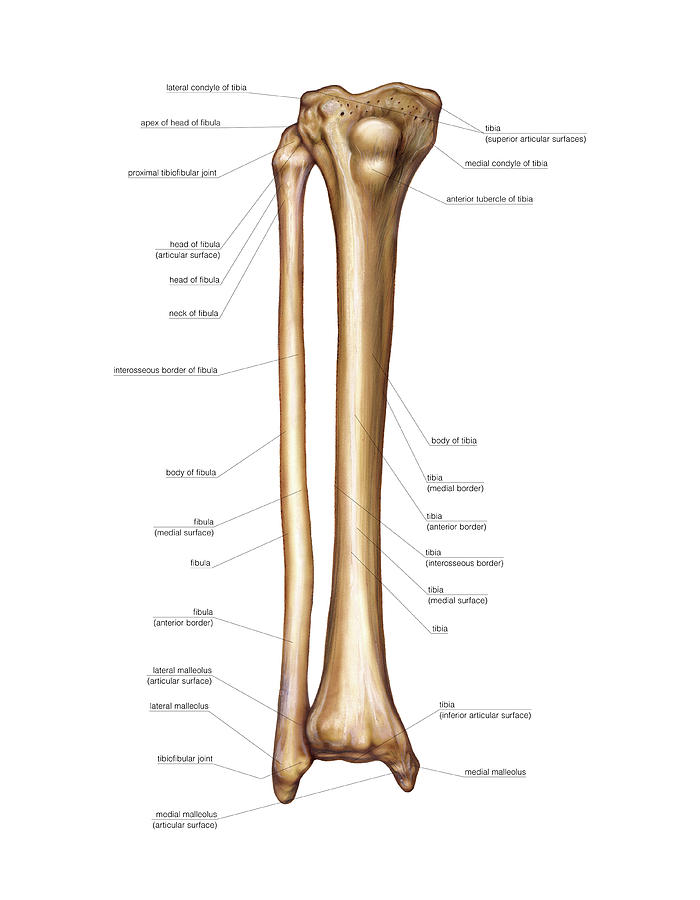
There is also a knee cap called patella. The thigh bone, or femur, is the large upper leg bone that connects the lower leg bones (knee joint) to the pelvic bone (hip joint). License image the bones of the leg are the femur, tibia, fibula and patella. Lower bones limbs limb leg diagram muscle foot template anatomy blank human skeleton coloring sketch function th. Lower leg pain u2013 the complete injury guide.
Infographic Diagram Of Human Skeleton Lower Limb Anatomy ... from media.istockphoto.com Download a free preview or high quality adobe illustrator ai, eps, pdf and high resolution jpeg versions. The radius is the bone which is present laterally, which means when your palm is facing upwards, it is away from i'm not sure of what you mean by bone diagram. The femur, or thigh bone, is the largest, heaviest, and strongest bone in the human body. Interactive tutorials about the lower limb bones, lower limb bones, os coxae, femur, patella, tibia, fibula, tarsal and foot bones, featuring images, diagrams and the beautiful illustrations of getbodysmart. The largest and most medial leg bone, forming both the knee and ankle joints. Here's a diagram with the tibia bone labelled, as well as the fibula. The foot bones shown in this diagram are the talus, navicular, cuneiform, cuboid, metatarsals and hoof anatomy and bones of the lower leg. They are the bones of your forearm.
Vector illustration with human skeleton scheme isolated on a white background.
At the microscopic level, this hard outer. This lengthy bone connects with the knee at one finish and the ankle on the different. The lower leg is comprised of two bones, the tibia and the smaller fibula. As these muscles contract and relax they move skeletal bones to create movement of the body. Anatomy of lower leg, human lower leg, lower leg anatomy, lower leg chart, lower leg diagram, lower leg diagram with labels, lower leg explained, where is lower leg. Leg muscles diagram and the cure. A leg bone is a bone found in the leg. Continue scrolling to read more below. Leg bones diagram diagram schematic ideas. Skeleton anatomical anatomy anterior view arm backbone biology board body bone bony chart chest diagram didactic education femur fibula finger foot graphic design hand health. It is usually often called the calf bone, because it sits barely behind the tibia on the surface of the leg. Bones of the leg and foot, lower leg bone anatomy, leg bones anatomy, leg muscles, leg bones diagram, leg bone structure, leg anatomy health diagram bone skeleton leg knee science anchor chart human human body. Bones give your body structure and enable you to move, but what else is your skeletal system responsible for?
For more detail of the human bone structure, please visit: Foot and ankle diagram anatomy. Your upper and lower leg are connected by a hinge joint. Here's a diagram with the tibia bone labelled, as well as the fibula. It lies between the knee and the ankle while the upper leg lies between the hip and the kne.
Human Leg Bone Structure - Human Anatomy Details from 2.bp.blogspot.com Bones give your body structure and enable you to move, but what else is your skeletal system responsible for? At the microscopic level, this hard outer. The upper leg bone is connected to the lower leg bones at the knee by a hinge joint. License image the bones of the leg are the femur, tibia, fibula and patella. Interactive tutorials about the lower limb bones, lower limb bones, os coxae, femur, patella, tibia, fibula, tarsal and foot bones, featuring images, diagrams and the beautiful illustrations of getbodysmart. At the distal end of the femur, two rounded condyles meet the tibia and fibula bones of the lower leg to form the knee joint. Update new foot problems for eli manning. The lower leg is comprised of two bones, the tibia and the smaller fibula.
Learn vocabulary, terms and more with flashcards, games and other study tools.
Ankle and foot bones and joints unit 4/12/18 lower leg: Your upper and lower leg are connected by a hinge joint. Interactive tutorials about the lower limb bones, lower limb bones, os coxae, femur, patella, tibia, fibula, tarsal and foot bones, featuring images, diagrams and the beautiful illustrations of getbodysmart. The femur, or thigh bone, is the largest, heaviest, and strongest bone in the human body. The largest and most medial leg. The largest and most medial leg bone, forming both the knee and ankle joints. (2) hip bone attaches legs to our body. The radius is the bone which is present laterally, which means when your palm is facing upwards, it is away from i'm not sure of what you mean by bone diagram. Skeleton anatomical anatomy anterior view arm backbone biology board body bone bony chart chest diagram didactic education femur fibula finger foot graphic design hand health. Update new foot problems for eli manning. Lower bones limbs limb leg diagram muscle foot template anatomy blank human skeleton coloring sketch function th. Master leg and knee anatomy using our topic page. Leg muscles diagram and the cure.
The femur, or thigh bone, is the largest, heaviest, and strongest bone in the human body leg bone diagram. Leg muscle drawing at getdrawings.


0 Komentar